# RKS: WAT - THE OBESITY SIGNAL - Understanding The Bulge-Causing Adipose Tissue
# RKS: WAT - THE OBESITY SIGNAL
- UNDERSTANDING THE BULGE-CAUSING ADIPOSE TISSUE
1st June 2022
WAT - BAT - bAT
All About Burning & Browning Of WAT
Dear Reader,
Obesity means having a Body Mass Index (BMI) >30 kg/m2 which is derived by dividing weight in kg by square of height measured in meters. What increases the BMI is the quantum of fat tissue which rises from 20% of body's content in normal lean individuals to 30-44% in those overweight and obese.
Fat tissue is called adipose tissue and the key to loosing weight therefore lies in burning fats.
ADIPOSE TISSUE TYPES
- White Adipose Tissue (WAT): This stores fatty acids (fats) in the form of triglycerides.
- Brown Adipose Tissue (BAT): This burns fatty acids breaking down triglycerides. Subsequently the triglycerides of WAT break down to release free fatty acids (FFA) in blood and these FFAs are taken up by BAT for further burning.
- Beige or Brite or Brown-like Adipose Tissue (bAT): When adipocytes of WAT are converted to beige coloured cells these are called brite cells; the WAT will then display brown areas within white adipocytes which is now referred to as bAT.
WHITE vs BROWN ADIPOCYTES
The fat in WAT is actually yellow in humans but white in animals. The yellow color of so-called WAT is because humans can't quickly metabolize the yellow carotene found in vegetables and grains; mainly beta-carotene then migrates to white fatty tissue and stains adipocytes yellow.
The brown adipocytes owe their color to the presence of iron in mitochondria. In comparison to less than 10% mitochondrial content of WAT, the BAT is packed with these energy houses. Mitochondria are the engines which burn the triglycerides and convert the fat into energy and simultaneously generating heat (thermogenesis), as a by-product, to keep the body warm.
PRINCIPLES OF LOOSING WEIGHT
There are only 2 possibilities to lose weight ultimately:
- Convert WAT to bAT.
- Increase the burning of the BAT & bAT.
In adults, 20% of adipose tissue is BAT but in those more than 50 years of age, the quantity declines to less than 10%. It is present in select areas of neck and shoulder (47-87% of total BAT) as well as in abdomen (belly).
Since adults have a limited distribution of BAT, the best option is to ensure as much as possible browning of WAT and facilitate burning of fats by brown adipocytes.
WHY TARGET WAT?
- Metabolically Healthy Obesity (MHO): Individuals have significantly increased BMI but it is more scWAT related.
- Metabolically Unhealthy Obesity (MUHO): These are typically individuals having increased vWAT that causes cardiometabolic disorders which include insulin resistance (type 2 diabetes), high lipids (dyslipidemia), high blood pressure (hypertension) besides increased risk of clot formation (thrombogenic profile).
In MUHO, the white adipocytes first increase in size (hypertrophy) due to overfilling of WAT with dietary fats (3 fatty acid molecules get converted to 1 triglyceride molecule) and manifesting as overweight or obesity. Later on, these white adipocytes increase in number (hyperplasia), which is genetically determined, and is associated with diabetes and high cholesterol (dyslipidemia) – metabolic syndrome. Hence, especially in MUHO, inducing browning of WAT, i.e. ibAT formation, is not only desirable to lose weight but also to tackle the associated metabolic syndrome.
ATTACKING & BROWNING OF WAT - BURNING BAT / bAT
ATTACKING WAT
The prime function of WAT is to generate energy in the form of adenosine triphosphate (ATP) primarily by utilizing the glucose. Besides there are 2 hormones secreted by adipocytes that govern WAT and are responsible for metabolic syndrome associated with overweight or obesity:
- LEPTIN: Increase breakdown of fats is leptin driven. In the obese there is leptin resistance resulting in decreased triglyceride breakdown to FFA and thereby enhanced fat tissue (WAT) accumulation.
- ADIPONECTIN: This hormone is important to enhance insulin sensitivity and thereby prevent insulin resistance development which is causative of diabetes. Adiponectin becomes deficient as age advances and this is the reason for development of metabolic disorders.
Hence, decreased adiponectin secretion by WAT causes obesity-associated complications. Also macrophages, defense cells type, are 2- to 4-fold higher in vWAT as compared to scWAT. These cells liberate inflammation causing chemicals (mediators) and the most important is tumor necrosis factor-alpha (TNFα). The persistent inflammation especially due to high levels of TNFα is the cause of developing insulin resistance in MUHO.
Approximately 60 to 85% of the weight of white adipose tissue is lipid, with 90-99% being triglyceride. Developing leptin resistance by the elderly thus causes obesity since these triglycerides in WAT cannot be broken down.
If obesity is to be tackled, the hormones secreted by WAT must be normalized, and the inflammatory mediators elaborated by white adipocytes need also to be reduced.
BROWNING OF WAT
Since presence of excessive WAT is the culprit in causing obesity, the conversion of white adipocytes into beige - brown-like adipocytes is another attractive option in management of overweight. Browning entails swarming the white adipocytes with mitochondria which will cause heat generation by burning fatty acids with consequent replenishing from stored triglycerides in WAT in an effort to supply more FFA to BAT.
The mitochondria contains peroxisome proliferator-activated receptor-gamma (PPAR-γ) coactivator 1α (PGC-1α), and PGC-1α is considered the master regulator of browning. PGC-1α activates PPAR-γ which then binds to the deoxyribonucleic acid (DNA) of adipocyte. Messenger ribonucleic acid (mRNA) sent by DNA releases enhancers which bind to PPAR-γ and promotes expression of UCP1 (uncoupling of protein-1) genes. Presence of UCP1 signals browning of WAT. PGC-1α promotes mitochondria multiplication and UCP1 facilitates the thermogenesis within the BAT mitochondria.
If ibAT conversion from WAT is to be facilitated, both PGC-1α and UCP1 need to be enhanced.
BURNING OF BAT / ibAT
The BAT, in contrast to WAT, is more richly supplied by blood vessels and nerves (sympathetic subtype of autonomous nervous system). When the sympathetic nerve is stimulated there is difference in responses in WAT and BAT / ibAT.
In WAT sympathetic nerve stimulation releases a chemical (neurotransmitter) called noradrenaline (NA). When NA binds with its receptor (adrenergic) the enzyme adenyl cyclase is activated and this breaks down ATP to cyclic adenosine monophosphate (cAMP). cAMP stimulates protein kinase (an enzyme) activity (PKA) – to form AMPK, to break down triglycerides in WAT.
In BAT the sympathetic nerve stimulation directly stimulates UCP1 which triggers AMPK and activate the SIRT1 gene. Both AMPK & SIRT1 stimulate PGC-1α (AMPK-SIRT1-PGC-1α pathway) which kickstarts the mitochondrial powerhouse. However, the protons required for ATP production are diverted by UCP1 from the energy processing and utilized to burn the released FFAs from WAT.
If burning of FFA by BAT / ibAT is to be enhanced, AMPK/SIRT1/PGC-1α pathway should be stimulated for thermogenesis is to be facilitated.
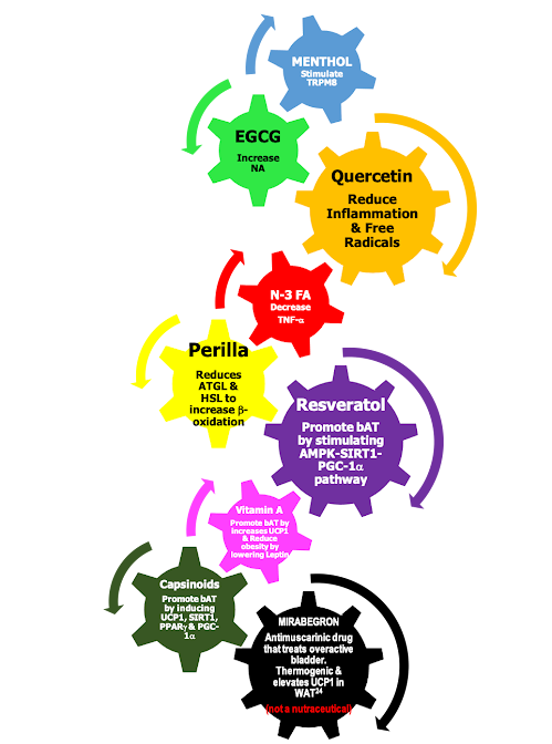
FACTORS STIMULATING BAT BURNING (THERMOGENESIS) & WAT BROWNING (bAT)
NUTRACEUTICALS FOR OBESITY
- Inflammatory mediators should be reduced to tackled the persistent state of inflammation of WAT.
- Browning of WAT to be induced (ibAT formation).
- BAT / bAT burning has to be facilitated such that more FFAs are released by WAT for ATP generation and WAT quantity is reduced.
Thus, a judicious blend of permutation and combination of above ingredients can be very helpful to ensure bAT formation and burning of brown adipocytes resulting in WAT reduction - obesity curtailing - risk reduction of associated cardiometabolic disease.
SUMMARIZING
WAT is the culprit for obesity but BAT is healthy. However, in adulthood, only 1-2% is the content of BAT (and constituting 4.3% of total fat mass) - so the best logical option is to convert WAT to BAT by inducing its browning - ibAT.
Exposure to cold can burn BAT / bAT but this is induced via increased NA and hence temporary. Better option is to convert WAT to bAT by expressing UCP1 and induce its burning by stimulating the AMPK-SIRT1-PGC-1α pathway.
10% weight loss in those with BMI >30 kg/m2 can reduce the risk of cardiometabolic sequelae by 21%. An average obese weighs 100 kgs, and has 1.5 kgs of BAT. If the desirable 0.5-1 kg weight loss per week is to occur 3 kgs of BAT is required to be burned. Hence, less than 1 kg of ibAT needs formed and such small degrees of WAT browning in MUHO populations can have a dramatic impact on cardiometabolic health.
- The vWAT is first lost – hence reducing the risk of cardiometabolic disorders immediately.
- Then is lost the scWAT from face first followed by legs and arms.
- Last is the loss of belly fat.
DR R K SANGHAVI
Experience & Expertise: Clinician & Healthcare Industry Adviser


Comments
Post a Comment